
"We speak both English and Spanish."
How Often Should You Get Dental X-rays? Guidelines for Different Patients
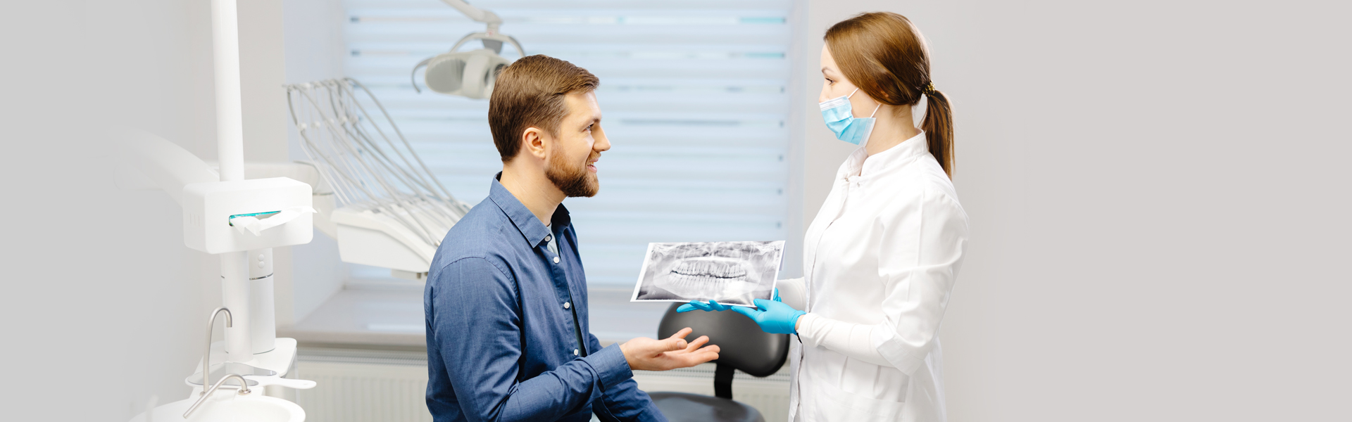
Dental X-rays play a vital role in dental treatments. They have allowed dentists to diagnose and treat oral issues such as decay and periodontal infections. It’s become customary for everyone to get a dental X-ray during a dental check-up examination. Visit our dentist at 94806 to learn more about different types of dental X-rays.
How X-ray Works and Types of X-Rays
X-rays are electromagnetic radiation that takes images or pictures of bones and soft tissues. They use a low and safe amount of radiation to achieve this. Medical practitioners often use X-rays to diagnose conditions and create customized patient treatment plans.
How X-rays Work
Dental X-rays can be taken from outside your mouth or inside while biting a metal plate. X-ray images are produced when high-velocity electrons collide with the metal plates and have energy which, together with X-rays, are absorbed by the metal plate.
A ray beam will travel through the air and come in contact with your body tissues or bone to produce an image film. The beam will pass through soft tissues like skin and organs that don’t absorb high-energy rays. Dense materials like bones absorb the radiation. Just like a camera, X-ray films will develop on areas that have been exposed to the X-rays. On the film, the white area will represent the denser tissues like bones, while the black areas will be the tissues that the rays have passed through, which are the soft tissues.
Types of Dental X-rays
Here are different X-rays used to provide information on various dental conditions.
Intraoral X-rays (inside the mouth)
- Bitewing. Bitewing X-rays are taken for preventative reasons and to detect decay on your lower and upper teeth. They capture the exposed part of the teeth(crown) and part of the supporting bone. You will be provided with an X-ray film to bite on if your dentist uses traditional X-rays, while digital X-rays will use a sensor that directs the X-ray to the computer for review.
- Periapical. These are taken to show the entire structure of your tooth and the jawbone. It’s used to find abnormalities in the surrounding bone and the root area.
- Occlusal. A large film will be placed against your lower or upper front teeth to capture the whole arch of your teeth, including the roof and the floor jaws. They check why your teeth haven’t yet erupted and show extra teeth. Occlusal X-rays are also used in diagnosing fractures or cleft palate, cysts, or abnormal growths.
Extraoral X-rays (outside the mouth)
- Panoramic. This uses a special machine to take an image of your entire lower and upper teeth. They are usually recommended to people who suffer from frequent complications or have undergone major dental work in the past. Panoramic X-rays are also used to prepare orthodontic treatments such as braces.
- Cephalometric Projection. This is an X-ray done on one side of the entire head. It is used by orthodontics to display how your teeth and jawbone fit together to create a personalized treatment plan for your whole mouth.
- Cone Beam X-ray. This type of X-ray uses an imaging method with computerized technology that converts two-dimensional images to three-dimensional pictures. The three-dimensional graphics can display every part of your tooth structure, bones, nerves, and soft tissues. Cone beam X-rays evaluate and plan complex oral surgery, such as implant placement and other dental procedures requiring precise and clear imaging.
Which is a Suitable Option for You
The condition of your dentition and treatment history determines the right dental X-ray option for you. Here are a few guidelines when selecting the proper digital X-rays in San Pablo, CA.
- If you are a new patient at the dental office, a complete mouth series of X-rays will be taken. Additionally, a panoramic may be recommended if you have malpositioned or impacted wisdom teeth.
- Suppose you are an existing patient returning for your routine visit. In that case, the dentist may recommend bitewing or full mouth X-rays if you have minimal caries, restorations, and healthy gum tissue for patients with a significant history of dental caries, root canals, or periodontitis. Panoramic X-rays will be taken for a general examination, while cone beam X-rays will be recommended after one to two years, depending on the risk of dental issues.
Are you looking for dental preventive measures to ensure your mouth is in optimal health? At iSmile San Pablo, we offer dental X-ray imaging during your dental examination to detect deformities, infections, and growths. Contact our dental office near you for more information.